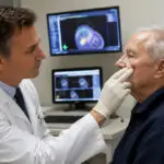
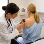
Your Strength. Our Knowledge and Experience.

When faced with a cancer diagnosis, the journey ahead is profoundly influenced by the support of the best doctors, options, and support team who work with you.

Mother with her two daughters
icon-5

We see you. All of you.

At El Portal Comprehensive Cancer Centers (EPCC), you will benefit from a dedicated team of doctors, nurses, and treatment professionals united by one common goal: an unwavering commitment to your health and well-being. 

icon-2

Your fight is personal for us.

We will deliver personalized, world-class care infused with compassion and kindness. Here, you can trust that you will receive cutting-edge treatment along with the highest quality of care.

Compassionate care is premiere at El Portal Cancer Center in the Central Valley of California
icon-4

Choose El Portal Comprehensive Cancer Centers as your trusted partner in this journey.

We are a multidisciplinary practice in Central California dedicated to providing care for patients with cancer and diseases of the blood.

Infusion Services

Our Infusion Center prioritizes your health with a commitment to providing excellent care. The first step towards better health is just a drip away, ensuring that you receive the support and treatment you need to enhance your well-being. Learn More information about Infusion Therapy.

Hematology/Oncology

Chemotherapy, Targeted Therapy, Immunotherapy, Iron (Infed, Venofer, Injectafer,  Feraheme, Monoferric), Therapeautic Phlebotomy

Neurology

IVIG, Tysabri, Solu-Medrol, Ocrevus, Leqembi,  Briumvi, Kisunla

IV Infusion/Injectable

Normal Saline (IV Fluids), IV Antibiotics, Prolia, Reclast, Evenity, Dupixent, & Xolair

Rheumatology

Rituxan, Actemra, & Orencia

Gastroenterology

Remicade, Entyvio, Stelera, Cimzia, & Skyryzi

Wellness

Vitamin C infusions, Myers Cocktail infusions,   Glutathione infusions, NAD+ Booster Infusions,  Inflammation and Detoxifying Treatments, Immune  Boosting Drip Treatments, Skin Rejuvenation   Infusions, as well as B12, Lipo-B, and NAD+   Injections

What our patients have to say...

april B. profile pictureapril B.
21:29 22 May 25
I feel fortunate to have been referred to Dr.M as my hematologist. He's been very attentive, thorough, and caring. He is a great Doctor who cares about his patients. Thank you so much Dr. M for caring, as well as to his staff who've been professional and kind also.
Stella Z. profile pictureStella Z.
15:40 19 May 25
Amazing place, front desk and doctors are friendly.
Neveah profile pictureNeveah
18:55 01 May 25
i love it here the ladies here are so nice and have a gentle hand when it comes to the needle. i feel comfortable and unjudged here and enjoy the time i spend here getting to talk to everyone.
Jaimie C. profile pictureJaimie C.
22:43 30 Apr 25
Love every single one of the girls who work in the infusion room. They do such a great job and always make sure we are comfortable and taken care of.
Natasha G. profile pictureNatasha G.
16:32 24 Apr 25
I can’t say enough great things about this place! From the moment I walked in, the staff made me feel more like family than a patient. Everyone is incredibly friendly, professional, and truly goes above and beyond to make sure you're comfortable. They explain every step of the process in detail, which really helps put your mind at ease.

The iron infusion room is spotless—super clean and sanitized, which I really appreciated. Danica and her amazing team are simply the best. Their care, compassion, and warmth made such a difference during my treatment. Once I’m done, I know I’ll truly miss them.

Thank you to the entire staff for making this experience as smooth and comforting as possible. Highly recommend!
Patricia L. profile picturePatricia L.
21:35 21 Apr 25
Thank you Isabel MA for your amazing help, best communication and treatment I received here much lovely service here
Mychelle C. profile pictureMychelle C.
19:00 26 Mar 25
Danika is awesome!!! She always makes sure to make sure I am as comfortable as possible!! The girls are all amazing!!

Share Your Journey with Us!

At El Portal Comprehensive Cancer Centers (EPCC), we believe that every story matters. Your experiences, struggles, and victories can inspire others who are navigating their own cancer journeys. By sharing your story, you not only help build a supportive community but also empower others to find hope and strength.

Working together to explore treatment options and to create a personalized plan based on your diagnosis and lifestyle.

News | Articles | EVENTS

Skin Cancer Symptoms

The most critical warning sign of skin cancer is the appearance of a new spot on the skin or changes in an existing spot that seem abnormal. Following the A B C D E guidelines can help you identify the common signs of skin cancer:

  • Asymmetry:One half of a mole (or birthmark) does not match the other half.
  • Border:Edges are irregular, ragged, notched, or blurred.
  • Color:The color is uneven, often featuring different shades of brown or black, along with patches of pink, red, white, or blue.
  • Diameter:The spot is larger than 6 millimeters across (about the size of a pencil eraser).
  • Evolving:The mole is changing in size, shape, color, or elevation. 

Skin Cancer Diagnosis

Skin cancer is typically identified first by a healthcare professional during an examination of an unusual area on the skin. However, determining whether it is cancerous requires removal and subsequent testing, known as a biopsy. This is the only reliable method to confirm a skin cancer diagnosis.

You may have your biopsy performed in a dermatologist’s office or as an outpatient procedure in a hospital or clinic, depending on the size and location of the abnormal area. Local anesthesia is usually administered to ensure your comfort.

There are four common types of skin biopsies:

  • Punch Biopsy:A sharp, hollow tool is used to remove a circle of tissue from the abnormal area.
  • Incisional Biopsy:A scalpel is employed to remove a portion of the growth.
  • Excisional Biopsy:The entire growth and some surrounding tissue are removed with a scalpel.
  • Shave Biopsy:A thin, sharp blade is used to shave off the abnormal growth.


If there is a possibility of melanoma, dermatologists will generally avoid simply shaving or cauterizing the growth. Instead, an excisional biopsy will be performed, or if the growth is too large, a tissue sample will be collected.

Staging Skin Cancer and Melanoma

Upon receiving a biopsy confirmation of skin cancer, you will be referred to a specialized oncologist for further evaluation and care. Understanding the extent, or stage, of your cancer is crucial, whether you have melanoma or nonmelanoma skin cancer. Staging involves a meticulous assessment to determine how thick the tumor is and whether it has metastasized to other parts of the body. If the tumor is particularly thick, your doctor may recommend chest x-rays, blood tests, and imaging scans of the liver, bones, and brain.

Knowing the stage of your cancer enables your oncologist to:

  • Assess the seriousness of your cancer and your chances for survival
  • Formulate the most effective treatment plan for your unique situation
  • Identify clinical trials that may present additional treatment options


In some cases, your oncologist may need to examine your lymph nodes to accurately stage the cancer. Surgical procedures may be part of the treatment plan to remove cancerous lymph nodes, which can aid in managing the disease.

Staging Criteria
Staging is based on several key factors:

  • The size of the tumor
  • How deeply it has penetrated below the surface of the skin
  • Whether it has spread to nearby lymph nodes or other organs


Oncologists utilize Roman numerals I through IV to designate the cancer stage. Stage I indicates small tumors that are confined to their original site, while Stage IV signifies advanced cancer that has spread to distant sites in the body.


Nonmelanoma Skin Cancer Staging
The stages of nonmelanoma skin cancer are as follows:

  • Stage 0: The cancer is localized to the top layer of skin, classified as carcinoma in situ.
  • Stage I: The tumor measures 2 centimeters (approximately three-quarters of an inch) or smaller.
  • Stage II: The tumor exceeds 2 centimeters in diameter.
  • Stage III: The cancer has spread beyond the skin to underlying tissues such as cartilage, muscle, or bone, or to nearby lymph nodes, but not to distant locations.
  • Stage IV: The cancer has metastasized to other areas in the body.


Melanoma Staging
The staging for melanoma specifically is defined as follows:

  • Stage 0: Melanoma cells are confined to the outer layer of skin (epidermis) and have not invaded deeper tissues.
  • Stage I: In Stage I, the melanoma is considered thin:
    • The tumor is no thicker than 1 millimeter (1/25 inch) and may show signs of ulceration (a scraped appearance).
    • Alternatively, the tumor measures between 1 and 2 millimeters thick without ulceration, and melanoma cells have not spread to nearby lymph nodes.
  • Stage II: At this stage, the tumor is at least 1 millimeter thick:
    • It may be between 1 and 2 millimeters thick and present with ulceration.
    • Alternatively, the tumor may be greater than 2 millimeters thick, potentially with ulceration, but has not yet spread to nearby lymph nodes.
  • Stage III: This indicates that melanoma has spread to nearby lymph nodes or to tissues just outside the original tumor without affecting the lymph nodes.
  • Stage IV: At this advanced stage, melanoma cells have metastasized to other organs, lymph nodes, or distant skin areas.
  • Recurrent: Recurrent disease indicates that cancer has returned after treatment, either in the original site or in a new location within the body.

Skin Cancer Treatment Based on Type

There are various approaches to treating skin cancer, and your oncologist will work closely with you to determine the most effective combination of therapies based on the stage of cancer and your overall health. We at El Portal Comprehensive Cancer Centers are dedicated to providing you with comprehensive care tailored to your needs, ensuring you receive the support, guidance, and treatment necessary for your journey toward recovery.


Nonmelanoma Skin Cancer Treatments

In some cases, the entire nonmelanoma skin cancer may be removed during the biopsy, which means no further treatment is necessary. However, if additional treatment is required, your doctor will outline the available options.

There are six primary types of nonmelanoma skin cancer treatments, which may be used individually or in combination:

  1. Surgery
  2. Chemotherapy
  3. Photodynamic therapy
  4. Radiation therapy
  5. Biologic therapy
  6. Targeted therapy


Melanoma Skin Cancer Treatments

Melanoma treatment encompasses nearly all the same options as nonmelanoma skin cancer, with the exception of photodynamic therapy.


Skin Cancer Treatment Options

Your oncologist can provide detailed explanations of the following surgical techniques:

  • Wide-Local Excision (WLE): This common approach involves numbing the area and surgically removing the cancer along with a margin of surrounding healthy skin. The margin is examined microscopically to ensure all cancer cells have been excised, and the required margin size will depend on the tumor’s dimensions. In some cases, nearby lymph nodes may also be removed.
  • Mohs Surgery: This technique is particularly beneficial for skin cancer treatment. The surgeon numbs the area and removes thin layers of the tumor, examining each layer under a microscope for cancer cells. The process continues until no cancerous cells are detected, ensuring complete tumor removal while preserving as much healthy tissue as possible.
  • Electrodesiccation and Curettage: This method is ideal for small basal cell skin cancers. The doctor numbs the area, removes the cancer using a curette (a spoon-shaped surgical instrument), and then employs an electric current to control bleeding and eliminate any remaining cancer cells. This procedure tends to be quick and straightforward.
  • Cryosurgery: This option is suitable for patients who may not tolerate other surgical methods. Cryosurgery employs extreme cold, typically generated from liquid nitrogen, to treat early-stage or very thin skin cancer lesions. While effective, this approach may cause swelling and could damage nerves in the area, leading to temporary loss of sensation.
  • Laser Surgery: This technique involves using a focused beam of light to target and destroy cancer cells. It is primarily utilized for superficial growths confined to the outer layer of skin.
  • Skin Grafting: Occasionally, grafts are necessary to cover openings left by surgical removal. In this procedure, the surgeon numbs the area and removes a patch of healthy skin from another part of your body, such as the thigh, to cover the excised area. It is important to provide special care to the graft site during the healing process.
  • Lymphadenectomy: This surgical procedure may be employed in the case of melanoma, wherein lymph nodes are removed for evaluation. The removed tissue is examined under a microscope for cancer cells.
  • Sentinel Lymph Node Biopsy: During this procedure, the sentinel lymph node—the first node likely to receive cancer spread from the tumor—is surgically removed. A radioactive substance and/or blue dye is injected near the tumor site to track the flow to the lymph nodes. If no cancer cells are found in the sentinel node, additional nodes may not need to be excised, making this a useful technique within melanoma treatment plans.

Dr. Alireza Mahmoudieh, M.D.

Dr. Mahmoudieh has been a vital member of El Portal Comprehensive Cancer Centers since 2008, bringing over a decade of dedicated experience to patient care. Before joining our team, he served as the Director of the Prostate Brachytherapy Program at Kaiser Permanente in Sacramento and Roseville, demonstrating his leadership in advanced cancer treatments.

He earned his medical degree from the Albert Szent-Györgyi School of Medicine in Szeged, Hungary, in 1996. With a strong interest in treating head, neck, and breast cancers, Dr. Mahmoudieh specializes in cutting-edge techniques such as Image-Guided Radiation Therapy (IGRT) and Intensity-Modulated Radiation Therapy (IMRT). His expertise extends to treating cancers of the head and neck, lung, gastrointestinal tract, and prostate.

A committed professional, Dr. Mahmoudieh is a member of the American Society for Therapeutic Radiology and Oncology (ASTRO), reflecting his engagement with ongoing developments in cancer treatment and research. His compassionate approach and advanced skill set ensure that each patient receives personalized, state-of-the-art care.

Outside of his professional life, Dr. Mahmoudieh enjoys traveling with his son and daughter, as well as reading and playing racquetball, embracing a balanced and active lifestyle. His dedication to both his patients and his personal passions makes him a trusted partner in your journey toward health.

Dr. Osman Elhanafy, Ph.D

Dr. Elhanafy is a highly respected Associate Professor of Medical Physics and serves as the Chief Medical Physicist at El Portal Comprehensive Cancer Centers since July 2013. He earned his Ph.D. in Medical Physics from the University of Wisconsin, Madison—where he began his foundational training and completed his thesis—in 2013, building on his initial joining of the university in 1999.

 With specialized training in advanced radiation oncology techniques—such as Intensity-Modulated Radiation Therapy (IMRT), High-Dose Rate Brachytherapy (HDR), Stereotactic Radiosurgery (SRS), Total Body Irradiation (TBI), Quality Assurance, and Radiobiology—Dr. Elhanafy brings a wealth of expertise to patient care. His dedication to the field is evident through numerous published research articles, as well as awards recognizing his leadership in organizing conferences, training sessions, lectures, and workshops.

Beyond his impressive credentials, Dr. Elhanafy is deeply committed to advancing medical physics and ensuring the best possible outcomes for his patients. His compassionate approach, combined with his extensive knowledge, makes him a trusted partner in your cancer care journey.

Outside of his professional pursuits, he values spending quality time with his family, reflecting his genuine care and balance in life.

Dr. Harish Ahuja, M.D.

Dr. Ahuja is a distinguished, board-certified Medical Oncologist with extensive training in medical hematology. He has refined his expertise at renowned institutions such as Tata Memorial Hospital, UCLA School of Medicine, and Roswell Park, under the mentorship of esteemed hematologists including Dr. S.H. Advani, Dr. Michael Caligiuri, and Dr. M.J. Cline.

With over 25 years of experience in both hematology and oncology, Dr. Ahuja offers a deep well of knowledge and a compassionate approach to patient care at El Portal Cancer Center. His dedication to advancing the field is reflected through active membership in prestigious professional organizations, including the American Society of Hematology, the American Society of Clinical Oncology, the European Society of Medical Oncology, and the Indian Society of Medical and Pediatric Oncology.

A passionate researcher, Dr. Ahuja has contributed significantly to ongoing scientific advancements—serving on the NRG Breast Committee and the Rare Subtype Committee, and acting as Principal Investigator for the “CROWN” NCORP study. His commitment to clinical excellence and groundbreaking research underscores his dedication to improving outcomes for his patients.

Outside of his professional pursuits, Dr. Ahuja values spending quality time with his family, and enjoys hiking and traveling, embracing life outside the clinic with the same dedication he brings to his medical practice. His compassionate nature and extensive experience make him a trusted partner in your cancer care journey.

Dr. Sidney Crain, M.D.

Dr. Crain is a highly experienced and board-certified Medical Oncologist and Hematologist with over 45 years of dedicated service in the medical field. Throughout his distinguished career, he has established himself as a leading expert in the diagnosis, treatment, and prevention of various cancers and blood disorders.

He earned his medical degree from the University of Illinois College of Medicine in 1977 and is actively affiliated with esteemed institutions, including Adventist Health St. Helena. Dr. Crain’s extensive experience spans a broad range of conditions, including small intestine and colorectal cancers, breast cancer, prostate cancer, anemia, leukemia, and lymphoma.

Beyond his clinical expertise, Dr. Crain is deeply committed to advancing medical research and improving patient care. His approach to treatment is rooted in compassion and a thorough understanding of complex medical conditions, ensuring each patient receives personalized, comprehensive care.

Recognized for his empathetic nature and unwavering dedication, Dr. Crain strives to achieve the best possible outcomes for his patients and foster a trusting, supportive environment. His lifelong commitment to the fields of oncology and hematology reflects his passion for making a meaningful difference in his patients’ lives.

Michael Kangas

Michael Kangas serves as the Director of Oncology and Diagnostic Imaging, where he spearheads initiatives that enhance revenue, profitability, productivity, and patient satisfaction within an underserved area. His leadership is instrumental in developing and guiding a highly skilled team of healthcare professionals across multiple clinics, providing comprehensive cancer care and diagnostic imaging services utilizing the most advanced technology. Michael’s commitment is rooted in delivering personalized, high-quality care to each patient, ensuring they receive the support they need throughout their journey.

With extensive expertise in Health Information Management, Electronic Medical Records (EMR), Radiation Therapy, Radiation Oncology, Clinical Operations, Healthcare Compliance, and Regulatory Standards, Michael ensures that all services meet the highest industry standards. He also focuses on monitoring performance, healthcare marketing, and supervisory skills to foster a collaborative, efficient, and patient-centered environment.

In addition to his clinical leadership, Michael actively volunteers with the Merced Cancer Society Foundation as its Executive Director. In this role, he oversees key operational and administrative functions to advance the foundation’s mission of positively impacting residents in the Central Valley. His efforts include community outreach, organizing fundraising events, and leading initiatives to expand the foundation’s support and resources for local cancer patients.

Michael’s dedication to improving healthcare and strengthening community ties highlights his unwavering commitment to healing, hope, and making a meaningful difference in the lives of those he serves.

Paul Mosher

Paul I. Mosher is a highly accomplished business professional with extensive experience in healthcare management, medical device sales, and community service. Currently, he proudly serves as the Physician Liaison and Business Development Coordinator at El Portal Comprehensive Cancer Center. In this role, Paul collaborates closely with our dedicated medical team to facilitate patient-centered care, foster strong relationships with our physicians, and support the center’s mission of providing the highest quality cancer treatment.

With expertise in business development, leadership, relationship management, and healthcare technology, Paul is a dedicated and effective communicator committed to exceptional service and compassionate support. His collaborative approach helps ensure that patients receive not only cutting-edge treatment but also personalized care rooted in trust and understanding. Additionally, Paul brings ten years of non-profit experience to our board of directors, furthering his devotion to community health and service.

Paul holds a Bachelor of Business in Business Marketing from Boise State University. He is actively involved in his community, volunteering with various Merced non-profits and churches. Outside of work, Paul enjoys spending quality time with his family and playing golf whenever he can.

Anemia

Anemia is the most prevalent benign blood disorder, impacting more than 3 million Americans, according to the National Heart, Lung, and Blood Institute. Anemia occurs when there is a deficiency of red blood cells or hemoglobin in the blood.

Common Symptoms:

  • Weakness
  • Shortness of breath
  • Dizziness
  • Pale skin


Anemia can particularly affect women during pregnancy or their childbearing years due to increased iron demands.

Causes of Anemia:

  1. Iron Deficiency Anemia: The most common type, usually caused by blood loss from heavy menstrual cycles or gastrointestinal issues, or from inadequate dietary iron. Treatment typically involves iron supplementation, either orally or intravenously.
  2. Aplastic Anemia: A rare condition where the bone marrow fails to produce enough blood cells. Causes may include exposure to toxins, medications, or autoimmune disorders. Diagnosis often requires a bone marrow biopsy. Treatment may include immunosuppressive therapies or bone marrow transplantation.
  3. Hemolytic Anemia: Occurs when red blood cells are destroyed faster than they can be produced. Causes range from infections to autoimmune disorders, with treatment addressing underlying causes and sometimes using corticosteroids.


Anemia Treatment
depends on its cause, severity, and the individual’s overall health. Here are some common treatment options:

  1. Dietary Changes: Increasing intake of iron-rich foods (e.g., red meat, beans, lentils, spinach) and foods high in vitamin B12 and folate (e.g., dairy, eggs, leafy greens).

  2. Iron Supplements: Oral or intravenous iron supplements may be prescribed, especially for iron-deficiency anemia.

  3. Vitamin Supplements: If the anemia is due to vitamin B12 or folate deficiency, supplements or injections may be recommended.

  4. Medications: For anemia related to chronic diseases, medications such as erythropoietin-stimulating agents may be used to stimulate red blood cell production.

  5. Blood Transfusions: In severe cases, a transfusion of red blood cells may be necessary to quickly increase hemoglobin levels.

  6. Managing Underlying Conditions: Treating the underlying cause of anemia, such as chronic kidney disease, can alleviate symptoms.

  7. Bone Marrow Stimulants: In certain cases, treatments that stimulate the bone marrow to produce more red blood cells may be indicated.

It’s essential to consult a healthcare provider for a proper diagnosis and treatment plan tailored to the individual’s specific condition and needs.T

Bleeding Disorders

Bleeding disorders are conditions in which the blood does not clot properly, leading to excessive bleeding. At El Portal Comprehensive Cancer Centers, our experts specialize in treating various bleeding disorders, including hemophilia and von Willebrand disease.

  • Hemophilia: A genetic disorder mainly affecting males, characterized by excessive bleeding from minor injuries. Modern treatments use synthetic concentrated clotting factors administered intravenously, along with vasopressin to manage bleeding.
  • Von Willebrand Disease : An inherited condition caused by a deficiency in a protein essential for blood clotting. Treatment focuses on preventing and managing bleeding episodes, often using synthetic hormones similar to vasopressin.

Treatment options for bleeding disorders like hemophilia and von Willebrand disease (vWD) focus on managing and preventing bleeding episodes. Here are common approaches for each:

Hemophilia:

  1. Factor Replacement Therapy:
    • Prophylactic Treatment: Regular infusions of clotting factor concentrates (Factor VIII for Hemophilia A and Factor IX for Hemophilia B) to prevent bleeding episodes.
    • On-Demand Treatment: Administering clotting factors when a bleeding episode occurs.
  2. Desmopressin (DDAVP):
    • Used for mild cases of Hemophilia A to stimulate the release of stored Factor VIII from blood vessel lining.
  3. Gene Therapy:
    • Investigational treatments aim to provide a functional copy of the missing clotting factor gene, potentially offering a long-term solution.
  4. Supportive Treatments:
    • Pain management and rehabilitation for joint health, along with lifestyle adjustments to prevent injury.


von Willebrand Disease (vWD):

  1. Desmopressin (DDAVP):
    • The first line of treatment for many with type 1 vWD and some with type 2, as it helps increase von Willebrand factor (vWF) and Factor VIII levels in the blood.
  2. Factor Replacement Therapy:
    • Concentrates containing von Willebrand factor and Factor VIII may be used for severe cases or during surgery.
  3. Antifibrinolytic Agents:
    • Medications like tranexamic acid may be used to help prevent bleeding during dental procedures or other situations.
  4. Avoiding Medications That Affect Clotting:
    • Recommendations to avoid aspirin and non-steroidal anti-inflammatory drugs (NSAIDs) that can increase bleeding risk.
  5. Lifestyle and Preventive Measures:
    • Education on avoiding injuries and managing heavy menstrual bleeding in women.


It’s crucial for individuals with bleeding disorders to work closely with a healthcare provider or a specialized hematologist to tailor treatment plans based on their specific needs and to manage their condition effectively.

Blood Clotting Disorders

Treatment options for blood clotting disorders aim to prevent clots from forming, manage existing clots, and reduce the risk of complications. Here are some common approaches based on different types of clotting disorders:

1. Anticoagulant Medications:

– Warfarin (Coumadin): A vitamin K antagonist used to prevent the formation of new clots and to keep existing clots from growing.

– Direct Oral Anticoagulants (DOACs): Medications such as apixaban (Eliquis), rivaroxaban (Xarelto), and dabigatran (Pradaxa) that directly inhibit specific clotting factors.

2. Antiplatelet Medications:

– Aspirin: Often used to prevent clot formation in arteries, particularly in individuals with a history of heart attacks or stroke.

– Clopidogrel (Plavix): Another antiplatelet medication that may be used alone or in combination with aspirin.

3. Thrombolytics:

– Clot Busters: Medications like alteplase (Activase) are used in emergency situations to dissolve large clots quickly, such as those causing strokes or pulmonary embolisms.

4. Compression Therapy:

– Compression Stockings: Used to improve blood flow and reduce swelling in individuals with venous thromboembolism (VTE) or chronic venous insufficiency.

5. Surgery:

– Thrombectomy: Surgical removal of a blood clot may be necessary in severe cases.

– Placement of Inferior Vena Cava (IVC) Filters: A filter may be placed in the IVC to catch clots that may travel from the legs to the lungs, particularly in patients with recurrent clots.

6. Lifestyle and Preventive Measures:

– Dietary Changes: Monitoring vitamin K intake (for patients on warfarin) and maintaining a balanced diet.

– Regular Exercise: Encouraging activity that enhances blood flow, while ensuring safety to avoid injury.

– Hydration: Staying well-hydrated to help maintain proper blood viscosity.

7. Management of Underlying Conditions:

Treating conditions that contribute to clotting disorders, such as atrial fibrillation, hormonal imbalances, or certain types of cancer.

Genetic Counseling:

For hereditary clotting disorders, genetic counseling may be beneficial for affected individuals and their families to understand risks and management strategies.

It’s essential for individuals with blood clotting disorders to work closely with a healthcare provider to develop a tailored treatment plan and to monitor their condition effectively. Regular follow-up and education about the disorder are also crucial parts of effective management.

Myelodysplastic Syndromes (MDS)

Myelodysplastic syndromes represent a group of disorders where the bone marrow fails to produce healthy blood cells, leading to insufficient white blood cells, red blood cells, and platelets. Patients with MDS may experience:

  • Increased risk of infections due to low white blood cell counts.
  • Fatigue and shortness of breath from anemia.
  • Excessive bleeding due to low platelet levels.


Treatment options
may include blood transfusions, medications to stimulate blood cell production, or immune-suppressive therapies.

Types of Bladder Cancer

  1. Transitional Cell Carcinoma (Urothelial Carcinoma): The most common type, originating in the bladder’s inner lining.

  2. Squamous Cell Carcinoma: Less common, often linked to irritation and inflammation of the bladder.

  3. Adenocarcinoma: A rare form that develops from glandular cells in the bladder.

  4. Small Cell Carcinoma: A rare and aggressive type that resembles small cell lung cancer.

Bladder Cancer Diagnosis

  1. Medical History and Physical Examination: A healthcare provider will assess symptoms and perform a physical exam.

  2. Urinalysis: A urine test can reveal blood (hematuria) or abnormal cells.

  3. Cystoscopy: A thin tube with a camera is inserted into the bladder to visually inspect for tumors or abnormalities.

  4. Biopsy: If abnormal tissue is found, a biopsy may be conducted during cystoscopy to confirm the presence of cancer.

  5. Imaging Tests: CT scans, MRIs, or ultrasounds may be used to evaluate the extent of the disease.

Bladder Cancer: Signs, Symptoms, and Risks

Signs and Symptoms

  • Blood in Urine (Hematuria): The most common symptom.
  • Frequent Urination: A strong urge to urinate more often than usual.
  • Painful Urination: Discomfort or burning sensation while urinating.
  • Lower Abdominal Pain: Discomfort in the bladder or pelvic area.
  • Back Pain: Pain in the lower back may also occur in advanced stages.

Risk Factors

  1. Smoking: The leading risk factor associated with bladder cancer.
  2. Chemical Exposure: Contact with certain chemicals, especially in industrial settings.
  3. Chronic Irritation: Repeated urinary tract infections or bladder inflammation.
  4. Family History: A genetic predisposition can increase risk.
  5. Age and Gender: More common in older adults and men.

Bladder Cancer: Potential Treatment Options

This information provides a snapshot of bladder cancer, covering essential aspects for better understanding.

  • Surgery:
    Transurethral Resection (TURBT): A common procedure to remove tumors.
    Cystectomy: Removal of part or all of the bladder in advanced cases.
  • Chemotherapy:
    Used to kill cancer cells or prevent them from growing, either before or after surgery.
  • Immunotherapy:
    Treatments that help the immune system recognize and attack cancer cells.
  • Radiation Therapy:
    Can be used in conjunction with other treatments or for patients who cannot undergo surgery.
  • Targeted Therapy:
    Newer drugs target specific characteristics of cancer cells to block their growth.

How Does Radiation Therapy Work?

During radiation therapy, a team led by a radiation oncologist—a specialist in this treatment—will carefully plan and supervise your treatment. Each session typically involves directing high-energy radiation toward the cancer cells using specialized equipment. This targeted exposure fractures the DNA within the cancer cells, making it difficult for them to grow and multiply. While healthy surrounding cells may also be exposed to some radiation, most can recover and continue functioning normally, whereas cancer cells generally cannot.

At El Portal Comprehensive Cancer Centers, we utilize the latest and most advanced radiation technologies to minimize exposure to healthy cells. Our accredited equipment undergoes daily checks to ensure the highest level of precision in treatment.

Types of Radiation Therapy

There are two main categories of radiation therapy:

External Beam Radiation Therapy

This method delivers radiation from a machine outside the body, directing beams specifically at the cancer site. The equipment does not physically contact the body and can be adjusted to deliver radiation from multiple angles. Treatment typically occurs five days a week for a duration ranging from 1 to 10 weeks.

Technologies may include:

  • TrueBeam Radiotherapy:Offers advanced options tailored to your specific anatomy and cancer type.
  • Image-guided Radiation Therapy (IGRT):Uses imaging (such as CT or ultrasound) to accurately direct radiation.
  • Intensity-modulated Radiation Therapy (IMRT):Shapes radiation beams around tumors while sparing surrounding healthy organs.
  • Stereotactic Radiosurgery (SRS):A non-invasive approach treating irregularly shaped tumors, particularly in the brain.
  • Stereotactic Body Radiation Therapy (SBRT):Similar to SRS, but designed for tumors outside the skull.
  • Hypofractionated Radiation Therapy:A specialized technique for breast cancer treatment, depending on individual patient factors.


Internal Radiation Therapy

Internal radiation therapy, or brachytherapy, involves placing radioactive material directly inside or near the tumor. This may include various implants such as pellets, seeds, or tubes, which are inserted using minimally invasive methods.

At El Portal Comprehensive Cancer Centers, we offer:

  • High-Dose Rate (HDR) Brachytherapy:Often utilized for uterine or cervical cancers, this approach involves placing radioactive material for short durations multiple times.
  • Selective Internal Radiation Therapy (SIRT):An innovative, non-surgical procedure for liver tumors, delivered via tiny radioactive beads inserted directly into blood vessels leading to the tumor.

How Does My Doctor Determined If Radiation Therapy Is Right for Me?

After a cancer diagnosis, you will likely be referred to a cancer specialist, such as a surgical oncologist or medical oncologist. Your treatment plan, which may include radiation therapy, is tailored uniquely based on:

  • Type and grade of cancer
  • Size and location of the tumor
  • Sensitivity of surrounding tissue
  • Genetic components
  • Family history
  • Age, overall health, and medical background
  • Personal treatment goals and preferences


Your doctor will also consider whether you have previously received radiation to that area, as each body part has a safe lifetime dose of radiation. The decision to proceed with radiation for recurrence depends on the location of the tumor, cancer type, and past radiation exposure.

Radiation therapy may be recommended as a standalone treatment or in conjunction with surgeries, chemotherapy, or immunotherapy. At El Portal Comprehensive Cancer Centers, we typically can schedule your first appointment within two days of your referral.

Choosing El Portal Comprehensive Cancer Centers for Radiation Therapy

Radiation therapy demands a high degree of skill, precision, and experience. Selecting a provider with state-of-the-art technology and strict protocols ensuring equipment accuracy is essential.

At El Portal Comprehensive Cancer Centers, we have been delivering high-quality radiation therapy for patients across California for over 15 years. Our board-certified radiation oncologists bring expertise and compassion to your care.

Key factors to consider include:

  • Technology:Choose a center equipped with the most advanced equipment for optimal treatment.
  • Experience:Our dedicated team ensures patients receive knowledgeable and compassionate care.
  • Affordability:As the largest community-based cancer center, we strive to provide services in a cost-effective

Accepted Insurance Carriers

AUTHORIZATION REQUIRED: 

  • Alignment Health Plan    
  • AllCare IPA Merced (HMO/NO DERM)  
  • ALLIANCE (CCAH)
  • Anthem MediCAL Managed Care Prog.  
  • Brand New Day-Universal Care, Inc.- Medicare 
  • Calviva Health 
  • Community Care IPA
  • California Forensic Medical Group, Inc  
  • Health Plan of San Joaquin HMO
  • Kova Healthcare, Inc. IPA  
  • Imperial Health Medical
  • MediCAL  
  • Central Valley Medical Providers IPA
  • Sequoia Health IPA 
  • United Healthcare (AARP)
  • Sante Community Physicians  
  • Tricare TriWest Healthare
  • Alliance  
  • Western Growers Assurance Trust  
  • Central Valley Pace County Medical Services Program 
  • Esperanza IPA- Medicare HMO
  • Choice Program
  • HealthNet CA- First Health Hill Physicians 
  • Humana LaSalle Network Management IPA  
  • Meritage Medical Network
  • Seven Corners 


NO AUTHORIZATION REQUIRED: 

  • Aetna
  • Anthem
  • Blue Cross of CA  
  • Blue Shield of CA
  • Medicare 
  • UMR ACPN Network-America’s Choice PN  
  • First Health Network-Cofinity Galaxy Health Network
  • HealthSmart 
  • CIGNA
  • Anthem ACA Plans- Pathway EPO, PPO 
  • BS of CA Promise Health Plan
  • Medicare
  • Railroad Multiplan/PHCS  
  • NaphCare NX Health Network LLC d/b/a Valenz Access 
  • Provider Network of America (PNOA)
  • Wellcare
  • WelbeHealth 

What You Need to Do

  • Verify benefits, plan limits, referral, and pre-authorization requirements before your appointment.
  • Bring your insurance card(s).
  • Notify us of any insurance or personal information changes immediately.
  • Understand your financial responsibility for co-pays, deductibles, and non-covered services.

How EPCC Assists You

  • We verify and review your benefits.
  • We help obtain necessary referrals and authorizations.
  • We provide cost estimates.
  • We inform you about financial assistance options.
  • We bill your primary and, if authorized, secondary insurance.
  • For Medicare patients, supervising physician info will be included on your claims.
  • Our Financial Counselors can provide resources for you to help navigate obtaining traditional insurance coverage 

Your Responsibilities

  • Present your current insurance card at each visit.
  • Keep us updated on insurance info.
  • Pay co-pays at the time of service.
  • Cover any balances remaining after insurance payments.

How Multiple Myeloma is Diagnosed

Diagnosing Multiple Myeloma typically involves several steps:

  1. Medical History and Physical Examination:
    • Your healthcare provider will start by reviewing your medical history, discussing symptoms you’ve experienced, and conducting a thorough physical examination to check for signs of the disease, such as bone tenderness or swelling.
  2. Blood Tests:
    • Blood tests are essential in diagnosing Multiple Myeloma. Common tests include:
      • Complete Blood Count (CBC): Monitors levels of red blood cells, white blood cells, and platelets.
      • Blood Chemistry Tests: Assess kidney function and other vital parameters.
      • Serum Protein Electrophoresis (SPEP): Identifies abnormal proteins (monoclonal proteins) produced by myeloma cells.
      • Free Light Chain Assay: Measures the levels of free light chains in the blood and can indicate the presence of myeloma.
  1. Urine Tests:
    • A 24-hour urine collection may be performed to check for Bence Jones protein, a type of light chain protein released by myeloma cells.
  2. Bone Marrow Biopsy:
    • A definitive diagnosis often requires a bone marrow biopsy, where a small amount of bone marrow (usually from the hipbone) is extracted and analyzed for abnormal plasma cells.
  3. Imaging Tests:
    • Imaging techniques such as X-rays, CT scans, or MRIs are used to assess bone damage or lesions caused by myeloma. These tests help identify areas of bone weakening or fractures.

Causes & Risk Factors of Multiple Myeloma

While the exact cause of Multiple Myeloma remains unknown, several factors may increase the risk:

  1. Age: The risk of developing Multiple Myeloma increases with age, most commonly affecting individuals over 65.
  2. Gender: Men are more likely to develop Multiple Myeloma than women.
  3. Race: African Americans have a higher incidence of Multiple Myeloma compared to Caucasian populations.
  4. Family History: A family history of Multiple Myeloma or related plasma cell disorders may increase individual risk.
  5. Certain Medical Conditions: Conditions such as monoclonal gammopathy of undetermined significance (MGUS) or other blood disorders can predispose individuals to myeloma.
  6. Exposure to Chemicals: Long-term exposure to certain chemicals, such as benzene, and agricultural chemicals, may contribute to the development of myeloma.

Signs & Symptoms of Multiple Myeloma

Recognizing the signs and symptoms of Multiple Myeloma is crucial for early intervention:

  1. Bone Pain: Persistent pain, particularly in the back, hips, or ribs, may indicate damage to the bones caused by myeloma lesions.
  2. Fatigue: Anemia, caused by reduced red blood cell production, often leads to fatigue and weakness.
  3. Frequent Infections: Compromised immune function due to myeloma can make patients more susceptible to infections.
  4. Unexplained Weight Loss: Patients may experience unexplained weight loss with no changes in their regular diet or physical activity.

Multiple Myeloma Treatment Options

Treatment for Multiple Myeloma is generally tailored to individual patient needs and the disease’s specific characteristics. Here are the primary treatment options:

  1. Chemotherapy: Chemotherapy remains a cornerstone of treatment for Multiple Myeloma. Regimens may include combinations such as:
    • Bortezomib (Velcade):A proteasome inhibitor that blocks protein degradation pathways in cancer cells.
    • Dexamethasone:A corticosteroid used in combination with other drugs to reduce inflammation and manage symptoms.
    • Lenalidomide (Revlimid):An immunomodulatory drug that enhances the immune response against myeloma cells.
  2. Targeted Therapy: Targeted therapies focus specifically on cancerous cells, minimizing harm to normal cells:
    • Monoclonal Antibodies:Medications like daratumumab (Darzalex) are designed to target specific proteins on myeloma cells, leading to their destruction.
    • Carfilzomib (Kyprolis):A newer proteasome inhibitor used in patients with relapsed or refractory myeloma.
  3. Immunotherapy: Immunotherapies use the body’s immune system to fight cancer:
    • Chimeric Antigen Receptor (CAR) T-Cell Therapy:This innovative approach involves modifying a patient’s T-cells to better recognize and attack myeloma cells. It has shown promise for some patients with difficult-to-treat myeloma.
  4. Stem Cell Transplant: For eligible patients, stem cell transplant can provide an opportunity for long-term remission:
    • Autologous Stem Cell Transplant:This involves harvesting the patient’s own stem cells, administering high-dose chemotherapy to eliminate cancer cells, and then reinfusing the healthy stem cells.
  5. Radiation Therapy: Radiation can be used to relieve pain or target specific areas affected by multiple myeloma, particularly in cases of localized disease or bone lesions.
  6. Supportive Care: Supportive care plays a vital role in managing symptoms and improving quality of life. This may include medications for pain relief, management of anemia, hydration support, and nutritional counseling.

Emotional Impact

A cancer diagnosis is a profound life event that impacts not only the individual diagnosed but also their entire family.

  • Anxiety and Fear:
    The uncertainty surrounding diagnosis, prognosis, and treatment outcomes can induce intense anxiety and fear among family members. Children, in particular, may struggle to comprehend the situation, leading to heightened worries about losing a parent or feeling responsible for the illness. Adults may worry about the future, their loved one’s health, or their own ability to handle caregiving responsibilities.
  • Guilt and Anger:
    Family members might experience guilt, feeling they should have detected the illness earlier or questioning whether their actions contributed to the situation. Anger can also surface—either directed at the illness, healthcare providers, or even oneself—potentially leading to emotional withdrawal or conflict within the family.
  • Helplessness and Powerlessness:
    The inability to “fix” the situation often leaves family members feeling overwhelmed. They may grapple with a sense of helplessness, unsure how to provide meaningful support or maintain normalcy, which can foster feelings of frustration or despair.
  • Risk of Mental Health Issues:
    Caregivers, especially spouses or primary caregivers, are at increased risk for depression, anxiety, and other mental health problems due to sustained emotional and physical stress. Recognizing early symptoms and seeking professional help can be vital in preventing more severe mental health consequences.
  • Changes in Family Relationships:
    The illness can lead to shifting roles—children taking on more responsibilities or a partner assuming new caregiving duties—potentially causing tension or resentment. Communication about these changes is essential to maintain trust and emotional intimacy.

Physical and Financial Impact

  • Disrupted Routines and Daily Schedules:
    Treatment appointments, hospital stays, and recovery periods can disrupt established routines, affecting work, school, and household management. Flexibility and proactive planning are crucial to minimize stress and ensure essential needs are met.
  • Financial Strain:
    The costs associated with cancer treatment (medications, therapies, travel expenses) can lead to significant financial hardship. Families may need to explore insurance options, financial assistance programs, or community resources to alleviate some of these burdens.
  • Impact on Children’s Well-Being:
    Children’s educational progress and social development may suffer due to disrupted routines, emotional distress, or increased household responsibilities. Providing age-appropriate information, maintaining routines where possible, and offering emotional support can help children cope better.
  • Physical Fatigue of Caregivers:
    The physical demands of caring—such as assisting with daily activities, managing medications, and providing emotional support—can lead to exhaustion, sleep disturbances, and physical health issues. Caregivers should seek respite care and prioritize self-care to sustain their health.

Other Considerations

By understanding and addressing the broad spectrum of physical, emotional, and practical impacts, families can develop a resilient approach to facing cancer together. Seeking support early, maintaining open communication, and prioritizing self-care can empower family members to navigate this challenging journey more effectively.

Effective Communication: Open, honest, and age-appropriate dialogues help family members process their feelings and clarify misunderstandings. Establishing regular family meetings or check-ins can facilitate emotional sharing and problem-solving.

Seeking Support and Resources: Families should not hesitate to reach out for support. This includes friends, extended family, support groupspecific to cancer patients and their families, and mental health professionals. Counseling can provide coping tools and emotional relief.

Individualized Support and Care: Recognize that each family member reacts differently based on personality, coping style, and relationship to the patient. Tailoring support—whether through counseling, support groups, or practical assistance—is critical to addressing these diverse needs.

Additional Strategies for Coping:

Education: Learning about the specific cancer type and treatment options can lessen uncertainty.

Advocacy: Helping family members become educated advocates within healthcare settings fosters confidence and ensures their needs are addressed.

Resilience Building: Engaging in stress-reduction activities, mindfulness, or hobbies can bolster emotional resilience.

Strategies

  1. Promote Open and Age-Appropriate Communication
    • Use honest language suitable for the child’s age to explain the illness without causing unnecessary fear.
    • Encourage questions and listen actively to family members’ concerns.
    • Establish regular check-ins to share updates and emotional responses.
  1. Enhance Emotional Resilience
    • Practice mindfulness, meditation, or relaxation techniques together as a family.
    • Engage in enjoyable activities that provide relief and normalcy, such as hobbies, outdoor walks, or creative arts.
    • Recognize and validate each family member’s feelings without judgment.
  1. Foster Practical Planning and Organization
    • Create a family calendar that includes treatment schedules, appointments, and important deadlines.
    • Develop a caregiving plan that distributes responsibilities fairly and includes contingency plans for emergencies.
    • Keep important documents accessible, such as insurance papers and medical directives.
  1. Self-Care for Caregivers and Family Members
    • Schedule regular breaks and time for personal activities to prevent burnout.
    • Prioritize sleep, nutritious eating, and physical activity whenever possible.
    • Seek peer support groups or counseling to share experiences and gain encouragement.
    1. Build and Utilize Support Networks
    • Reach out to friends, extended family, and community resources early.
    • Consider virtual support options if local groups are unavailable or if mobility is limited.
    • Involve trusted individuals in caregiving and emotional support roles to lighten the load.
  1. Stay Informed and Advocate
    • Educate yourself about the specific cancer diagnosis, treatment options, and side effects.
    • Maintain open communication with healthcare providers to ask questions and express concerns.
    • Advocate for your family’s needs within the healthcare system to ensure comprehensive care.

Leukemia – Cause & Risk Factors

While the precise causes of leukemia remain unclear, certain factors have been identified that may increase the likelihood of developing the disease.

Known Risk Factors for Leukemia Include:

  • Genetic Factors: Individuals with certain genetic syndromes, such as Down syndrome, are at higher risk for leukemia. Specific inherited genes may also predispose individuals to developing the disease.
  • Environmental Exposure: Long-term exposure to high levels of radiation—such as that from nuclear accidents—or exposure to chemicals like benzene (found in cigarette smoke and industrial emissions) has been linked to an increased risk of leukemia.
  • Age: The incidence of leukemia increases with age, though acute forms can occur in children as well.
  • Family History: Individuals with a family history of leukemia or other blood cancers are at a higher risk, indicating a potential genetic component.
  • Previous Cancer Treatment: Those who have undergone chemotherapy or radiation for other cancers may have an elevated risk of developing leukemia later on.

Leukemia – Signs & Symptoms

Leukemia can present with a variety of symptoms, which may vary based on the type but commonly include:

  • Fatigue or Weakness: A significant drop in red blood cells can lead to anemia, causing chronic fatigue, dizziness, and weakness.
  • Frequent Infections: Abnormal white blood cells can interfere with the immune system, resulting in increased susceptibility to infections.
  • Easy Bruising or Bleeding: Low platelet counts can impair the blood’s ability to clot, leading to excessive bruising, nosebleeds, or gum bleeding.
  • Unexplained Weight Loss: Many patients experience unintentional weight loss as a result of the body’s heightened metabolic need in response to the disease.
  • Night Sweats: Some individuals experience drenching sweats at night, often accompanying fever and chills.
  • Pain in the Bones or Joints: Some forms of leukemia.

Leukemia – Diagnosis & Treatment

Diagnosis of Leukemia

Diagnosing leukemia typically involves a combination of medical history, physical examination, and specific tests, including:

  • Blood Tests:To look for abnormal levels of white blood cells, red blood cells, and platelets.
  • Bone Marrow Biopsy:A definitive test to confirm leukemia and identify its type.
  • Imaging Tests:Such as CT scans or MRIs, to assess any organ involvement.


Leukemia Treatment Options

Treatment for leukemia is tailored to the type and stage of the disease but may include:

  • Chemotherapy:The primary treatment for most types of leukemia, aiming to kill cancer cells.
  • Radiation Therapy:Used to target specific areas and relieve symptoms.
  • Targeted Therapy:Drugs that specifically target cancer cell growth signals.
  • Stem Cell Transplant:Harvesting healthy stem cells to replace diseased bone marrow.

Staging Leukemia

Staging is an essential process that helps determine the extent of the disease and guide treatment options.

Chronic Lymphocytic Leukemia (CLL) Stages

CLL is typically classified into the following stages:

  1. Stage 0: Low-risk; increased lymphocyte count without symptoms.
  2. Stage I: Enlarged lymph nodes.
  3. Stage II: Enlarged lymph nodes and spleen or liver.
  4. Stage III: Anemia due to low red blood cell counts.
  5. Stage IV: Low platelet counts and significant complications.


Rai Staging System

The Rai Staging System is a common method to categorize CLL, based on physical examination and blood test results, including the number of lymphocytes and the presence of anemia or low platelet levels.

Chronic Myeloid Leukemia (CML) Stages

CML has three phases:

  1. Chronic Phase: Slow growth, often asymptomatic at diagnosis.
  2. Accelerated Phase: Disease begins to evolve, with signs of progression.
  3. Blastic Phase: Rapid growth, resembling acute leukemia and requiring urgent treatment.

What to Expect at Your First Appointment

Medical History Review: The hematologist will ask detailed questions regarding your medical history, family history, and any symptoms you’ve been experiencing. Be prepared to provide information on previous illnesses, medications, and treatments you have received.

Physical Examination: A physical exam will often be performed to assess your overall health and identify any signs of hematologic disorders. This may include checking for signs of anemia, swelling in lymph nodes, or other related symptoms.

Diagnostic Tests: You may undergo blood tests or, in some cases, a bone marrow biopsy to determine the nature of your condition. If you already have recent test results, bring copies.

Discussion of Symptoms: You’ll have the opportunity to discuss any symptoms you have been experiencing, such as fatigue, infections, or bleeding. Be specific about the duration and severity of each symptom.

Treatment Plan Discussion: Based on your diagnostic results, the hematologist will discuss potential treatment options, including the benefits and risks associated with each.

Referrals and Additional Tests: If needed, you may be referred to other specialists, or additional diagnostic tests may be scheduled.

Questions to Consider Asking

When meeting with the hematologist, it’s essential to have your questions ready. Here are some key questions that can guide the conversation:

Diagnosis Clarification:

  • What specific type of hematologic condition do I have?
  • How was this diagnosis determined?


Implications of Diagnosis:

  • What does my diagnosis mean for my health?
  • Are there any associated risks or complications?


Symptoms and Management:

  • What symptoms should I watch for that would require immediate medical attention?
  • Are there any lifestyle changes I should consider to manage my condition?


Treatment Options:

  • What treatments are available for my condition, and which do you recommend?
  • What are the potential side effects of each treatment option?
  • How long will treatment last, and what does it involve?


Prognosis:

  • What is the prognosis for my condition?
  • How often will I need follow-up appointments or tests?


Support Resources:

  • Are there support groups or resources you recommend for patients with similar diagnoses?
  • How can I get in touch with a nutritionist or mental health professional if needed?


Questions Regarding Future Visits:

  • How frequently will I need to return for follow-ups?
  • What tests or evaluations will be conducted during future visits?


Additional Tips for Your Appointment

  • Bring a Support Person: Consider bringing a family member or friend for support. They can help take notes and remember information discussed during the appointment.
  • Prepare a List: Write down your symptoms, concerns, and any medications you are currently taking. This preparation can ensure that you don’t forget important details during the visit.
  • Be Honest: Feel free to share your emotional state or any fears you may have regarding your health. Hematologists are trained to address both physical and emotional aspects of care.
  • Follow-up Questions: Don’t hesitate to ask for clarification if you don’t understand something. It’s essential to have a clear understanding of your diagnosis and treatment plan.

By preparing thoughtfully for your first hematologic oncology appointment and asking the right questions, you’ll be better positioned to address your health concerns and collaborate effectively with your healthcare team.

Signs and Symptoms of Hodgkin Lymphoma

Hodgkin lymphoma (HL) can present with various signs and symptoms, which may evolve over time. Common symptoms include:

  1. Swollen Lymph Nodes: The most prominent sign is usually painless swelling of lymph nodes in the neck, armpit, or groin. These nodes may feel firm or rubbery.
  2. Fever: Patients may experience persistent fevers without an infectious cause, often referred to as “B symptoms.”
  3. Night Sweats: Drenching night sweats that can soak clothing and bedding, often severe and uncomfortable.
  4. Unexplained Weight Loss: Significant weight loss (more than 10% of body weight) over a short period without changes in diet or exercise.
  5. Fatigue: Persistent fatigue or lack of energy that does not improve with rest.
  6. Itching: Generalized itching (pruritus) can occur without a rash and may be bothersome.
  7. Chest Pain or Cough: If the lymphoma affects areas in the chest, individuals may experience chest pain, persistent cough, or difficulty breathing.
  8. Abdominal Pain or Swelling: Involvement of the abdomen may lead to pain or a sense of fullness due to enlarged lymph nodes or spleen.
  9. Alcohol-Induced Pain: Some patients report pain in lymph nodes after consuming alcohol, which is quite specific to Hodgkin lymphoma.

Risk Factors of Hodgkin Lymphoma

Several factors can increase the risk of developing Hodgkin lymphoma:

  1. Age: Hodgkin lymphoma is most commonly diagnosed in young adults aged 15-35 and older adults aged 55 and above.
  2. Gender: It is more common in males than females.
  3. Family History: A family history of Hodgkin lymphoma or other related cancers may increase risk.
  4. Weakened Immune System: Individuals with weakened immune systems, such as those with HIV/AIDS or those on immunosuppressive therapy, have an increased risk.
  5. Viral Infections: Infections with the Epstein-Barr virus (EBV) and human immunodeficiency virus (HIV) are associated with an elevated risk.
  6. Geographic Location: There are geographic variations in HL incidence, with higher rates reported in certain regions.
  7. Obesity: Some studies suggest a potential link between obesity and an increased risk of Hodgkin lymphoma.
  8. Previous Radiation Therapy: Individuals who received radiation therapy for other cancers, especially during childhood, may have a heightened risk.

Treatment Options for Hodgkin Lymphoma

The treatment of Hodgkin lymphoma (HL) often depends on the stage of the disease, tumor characteristics, the patient’s age, and overall health. Common treatment options include:

  1. Chemotherapy: Chemotherapy is the cornerstone of treatment for Hodgkin lymphoma. Common regimens include ABVD (doxorubicin, bleomycin, vinblastine, and dacarbazine), which is effective in most patients.
  2. Radiation Therapy: Radiation therapy may be used alone or in combination with chemotherapy, especially for localized disease (early-stage HL). It targets specific areas affected by lymphoma.
  3. Stem Cell Transplant: For patients with relapsed or refractory Hodgkin lymphoma, high-dose chemotherapy followed by autologous stem cell transplantation may be considered. This allows for the use of higher doses of chemotherapy to effectively destroy cancer cells.
  4. Immunotherapy: Newer targeted therapies, including monoclonal antibodies (like brentuximab vedotin), use the body’s immune system to fight cancer. This option may be used in certain cases of relapsed or refractory HL.
  5. Clinical Trials: Participation in clinical trials may provide access to innovative therapies and new treatment approaches that are not yet widely available.
  6. Supportive Care: Management of side effects and supportive care to maintain quality of life during treatment are crucial. This may include medication for nausea, pain management, and counseling services.

Statistics Regarding Treatment and Recovery

  1. Hodgkin Lymphoma: The overall 5-year survival rate for Hodgkin lymphoma is approximately 87%, with a significant number of patients achieving long-term remission. Cure rates in early-stage HL can be even higher, exceeding 90%with appropriate treatment.
  2. Advancements in Treatment: Cutting-edge treatments such as targeted therapies and immunotherapy (including CAR T-cell therapy) have shown promising results. In certain cases of relapsed/refractory NHL, CAR T-cell therapy has achieved response rates surpassing 50%.
  3. Chemotherapy Success: First-line chemotherapy regimens for Hodgkin and many types of non-Hodgkin lymphoma, such as R-CHOP (rituximab combined with chemotherapy), lead to high initial response rates, with many patients showing significant reduction or complete disappearance of tumors.
  4. Survivorship: Many lymphoma survivors go on to lead healthy, fulfilling lives. Long-term follow-up studies show that most individuals achieve a normal or near-normal quality of life after treatment, especially with supportive care and monitoring.


Treatment Modalities

  • Combination Chemotherapy: Most lymphomas are treated with combination chemotherapy, which enhances the effectiveness of treatment and reduces the risk of resistance. Such regimens have improved overall survival rates for various lymphoma types.
  • Radiation Therapy: For localized lymphoma, radiation therapy can be highly effective. It often leads to excellent outcomes, especially in early-stage Hodgkin lymphoma, with success rates over 90%.
  • Stem Cell Transplants: For patients with relapsed or refractory lymphoma, autologous stem cell transplants can offer a chance of long-term remission. Survival rates post-transplant can be around 50-60%, depending on various factors.


Ongoing Research and Future Prospects

Customized Treatment Approaches: Advances in genetics and molecular biology allow for more personalized treatment approaches, enabling physicians to tailor therapies based on individual patient characteristics and disease profiles.

Signs and Symptoms of Non-Hodgkin Lymphoma

Non-Hodgkin Lymphoma (NHL) can present with a variety of signs and symptoms, which may vary depending on the subtype of NHL and the areas of the body affected. Common signs and symptoms include:

  1. Lymphadenopathy: Swollen lymph nodes, particularly in the neck, armpits, or groin. These may feel rubbery or firm and are often painless.
  2. Fever: Persistent fevers without an obvious cause, sometimes with chills.
  3. Unexplained Weight Loss: Significant weight loss (more than 10% of body weight) without trying or without an underlying reason.
  4. Night Sweats: Drenching night sweats that may soak through clothing and bedding.
  5. Fatigue: Chronic fatigue or weakness that doesn’t improve with rest.
  6. Abdominal Pain or Swelling: Pain or discomfort in the abdomen, which may also be associated with a swollen abdomen due to enlarged lymph nodes or spleen.
  7. Chest Pain: If lymphoma affects the chest area, it can cause chest pain, coughing, or difficulty breathing.
  8. Itching: Generalized itching (pruritus) without a rash, which can be quite bothersome.
  9. Skin Changes: In some cases, NHL can present with skin rashes, lesions, or lumps.


It’s important to note that these symptoms can be caused by other health conditions as well; experiencing one or more of these symptoms doesn’t necessarily mean an individual has NHL. However, if symptoms persist or worsen, it is advisable to consult a healthcare professional for further evaluation.

Risk Factors of Non-Hodgkin Lymphoma

Several factors can increase the risk of developing Non Hodgkin lymphoma (NHL):

  1. Age: The risk of NHL increases with age, with most cases diagnosed in adults aged 60 and older.
  2. Gender: Men are more likely to develop NHL than women.
  3. Weakened Immune System: Individuals with weakened immune systems, such as those with HIV/AIDS, have a higher risk of developing NHL. Immunosuppressive treatments (e.g., for organ transplant patients) also increase risk.
  4. Autoimmune Diseases: Conditions such as rheumatoid arthritis, lupus, or Sjögren’s syndrome can increase the risk due to chronic inflammation and immune activation.
  5. Infections: Certain viral infections, such as the Epstein-Barr virus (EBV), human immunodeficiency virus (HIV), and human T-cell lymphotropic virus (HTLV), have been linked to an increased risk of developing certain types of NHL.
  6. Chemical Exposure: Exposure to certain chemicals, particularly pesticides, herbicides, and solvents such as benzene, may be associated with a higher risk of NHL.
  7. Family History: A family history of lymphoma or other blood cancers may increase an individual’s risk.
  8. Geographic Location: Some studies suggest that living in certain geographic regions with higher pesticide use or specific environmental exposures may contribute to an increased risk.
  9. Obesity: Some research indicates that obesity may be a risk factor for developing various types of lymphoma.
  10. Previous Radiation Therapy: Individuals who have received radiation therapy for other cancers may have an increased risk of developing NHL later.

Treatment Options for Non-Hodgkin Lymphoma

  1. Chemotherapy:Chemotherapy is often the first-line treatment for many types of NHL. It involves the use of powerful drugs to kill cancer cells or inhibit their growth. Commonly used chemotherapy regimens include:
    • R-CHOP:A combination of rituximab (a monoclonal antibody) with cyclophosphamide, doxorubicin, vincristine, and prednisone.
    • CHOP:Similar to R-CHOP but does not include rituximab.
  2. Targeted Therapy: Targeted therapies are designed to specifically attack cancer cells while minimizing damage to normal cells. Some examples include:
    • Rituximab (Rituxan):A monoclonal antibody targeting CD20-positive B-cell lymphomas.
    • Brentuximab Vedotin (Adcetris):A medication that combines an antibody with a chemotherapy drug, used for specific subtypes of NHL.
  3. Immunotherapy: Immunotherapy utilizes the body’s immune system to combat cancer cells. It includes therapies such as:
  4. CAR T-Cell Therapy:A groundbreaking treatment that involves modifying a patient’s own T-cells to target and destroy lymphoma cells, showing high effectiveness in certain types of aggressive NHL.
  5. Nivolumab (Opdivo) and Pembrolizumab (Keytruda):Immune checkpoint inhibitors that help the immune system recognize and attack cancer cells.
  6. Radiation Therapy: Radiation therapy may be used to target specific areas of lymphoma, especially for localized disease. It can be effective in combination with chemotherapy for certain patients.
  7. Stem Cell Transplant: For patients with high-risk or relapsed NHL, stem cell transplantation (autologous or allogeneic) may be considered. This approach allows for the use of high-dose chemotherapy to treat the lymphoma, followed by transplanting healthy stem cells to restore the bone marrow.
  8. Supportive Care: Throughout treatment, supportive care is essential to manage side effects and improve quality of life. This can include anti-nausea medications, pain management, nutritional support, and counseling services.
  9. Observation (Watchful Waiting): In some cases of indolent or slow-growing NHL, especially in asymptomatic patients, a strategy of “watchful waiting” may be recommended, where treatment is delayed until symptoms arise or the disease progresses.


Conclusion

Non-Hodgkin Lymphoma comprises a diverse group of diseases, and treatment plans are personalized based on many factors, including the specific lymphoma subtype and the patient’s overall health. Early diagnosis and access to effective therapies greatly enhance treatment success rates. It’s essential for patients to discuss all available treatment options with their healthcare providers to determine the most appropriate approach for their individual situation.

Statistics Regarding Treatment and Recovery

  1. Non Hodgkin Lymphoma:The 5-year survival rate for non-Hodgkin lymphoma varies widely based on the specific type and stage but is generally around 72%. For some aggressive subtypes, advances in treatment have improved survival rates markedly.
  2. Advancements in Treatment: Cutting-edge treatments such as targeted therapies and immunotherapy (including CAR T-cell therapy) have shown promising results. In certain cases of relapsed/refractory NHL, CAR T-cell therapy has achieved response rates surpassing 50%.
  3. Chemotherapy Success: First-line chemotherapy regimens for Hodgkin and many types of non-Hodgkin lymphoma, such as R-CHOP (rituximab combined with chemotherapy), lead to high initial response rates, with many patients showing significant reduction or complete disappearance of tumors.
  4. Survivorship: Many lymphoma survivors go on to lead healthy, fulfilling lives. Long-term follow-up studies show that most individuals achieve a normal or near-normal quality of life after treatment, especially with supportive care and monitoring.


Treatment Modalities

  • Combination Chemotherapy: Most lymphomas are treated with combination chemotherapy, which enhances the effectiveness of treatment and reduces the risk of resistance. Such regimens have improved overall survival rates for various lymphoma types.
  • Radiation Therapy: For localized lymphoma, radiation therapy can be highly effective. It often leads to excellent outcomes, especially in early-stage Hodgkin lymphoma, with success rates over 90%.
  • Stem Cell Transplants: For patients with relapsed or refractory lymphoma, autologous stem cell transplants can offer a chance of long-term remission. Survival rates post-transplant can be around 50-60%, depending on various factors.


Ongoing Research and Future Prospects

Customized Treatment Approaches: Advances in genetics and molecular biology allow for more personalized treatment approaches, enabling physicians to tailor therapies based on individual patient characteristics and disease profiles.